Glaucoma is a condition in which the normal intraocular pressure i.e. fluid pressure inside the eyes rises slowly.Glaucoma occurs when the fluid drainage from the eye is blocked by abnormal development or injury to the drainage tissues, thus, resulting in an increase in the intraocular pressure, damage to the optic nerve, and loss of vision.
Symptoms :
- light sensitivity (photophobia)
- corneal opacification (hazy gray cornea)
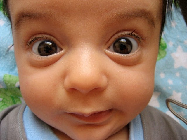
- enlarged eye and cornea
- epiphora (overflow of tears)
- vision loss
- cloudy cornea is the earliest and most common sign of childhood glaucoma.
- The healthy cornea is transparent and the loss of this transparency is caused by edema, or swelling of tissue from excess fluid. This occurs in the corneal epithelium (outermost layer of the cornea) and in the corneal stroma (middle layer of the corneal tissue).
- In most cases of glaucoma affecting children under three years of age, the cornea and eye enlarges.
- Secondary symptoms common with acute glaucoma include irritability, loss of appetite, and vomiting. These symptoms may be misunderstood before the glaucoma is recognized.
- young children with glaucoma are often unhappy, fussy, and poor eaters.
- there is discomfort and pain when the eye pressure increases rapidly during an acute onset or with the rapid return of glaucoma
Glaucoma is classified according to the age of onset. Glaucoma that begins before the child is 3 years old is called infantile or congenital (present at birth) glaucoma.

Signs to look out for all children under 18:
- child’s eyes particularly sensitive to sunlight or a camera flash,
- significant vision loss in your child
- child’s eyes have difficulty adjusting in the dark
- child complain of headaches and/or eye pain
- child blink or/and squeeze his/her eyes often
- child have red eyes all the time
Diagnosis

- Visual acuity test – the common eye chart test (with letters and images), which measures vision ability at various distances.
- Pupil dilation – the pupil is widened with eyedrops to allow a close-up examination of the eye’s retina and optic nerve.
- Visual field – a test to measure a child’s side (peripheral) vision. Lost peripheral vision may be an indication of glaucoma.
- Tonometry – a standard test to determine the fluid pressure inside the eye.
Management
- Medications: some medications cause the eye to produce less fluid, while others lower pressure by helping fluid drain from the eye.
- Surgery:The purpose of conventional surgery is to create a new opening for fluid to leave the eye.
surgery.Surgical procedures are performed by using microsurgery or lasers. The purpose of surgery is to create an opening for fluid to leave the eye.
- Trabeculotomy and goniotomy
A surgical opening is made into the drainage area of the eye therefore establishing a more normal anterior chamber angle that allows the fluid to drain more freely, lowering the intraocular pressur.
A goniotomy is an internal trabeculotomy procedure that is used in congenital glaucoma.
trabeculectomy.
A surgical procedure that involves the removal of part of the trabecular meshwork drainage system, allowing the fluid to drain from the eye. - Iridotomy
In this procedure, a small hole is made through the iris – the colored part of the eye – to allow fluid to flow more freely in the eye. The surgeon may use a laser to create this hole (laser iridotomy). - Cyclophotocoagulation
A procedure that uses a laser beam to freeze selected areas of the ciliary body – the part of the eye that produces aqueous humor – to reduce the production of fluid. This type of surgery may be performed with severe cases of childhood glaucoma
 +91-94 24 851 514